What Should I Know About the Knee Ligaments?
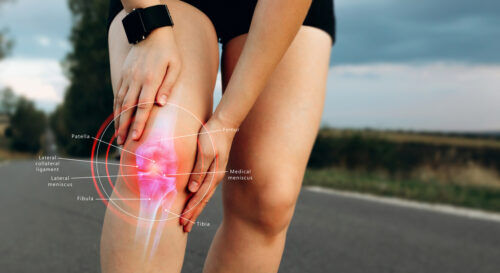
The quick answer is the knee ligaments provide stability to your knee joint. How many times have you been watching your favorite sporting event and heard an athlete that sprained a knee ligament, or even worse, tore it? Or, maybe you yourself have experienced some knee pain and don’t really know what to do about it? In this article, we will describe the anatomy and function of the knee ligaments.
Head, Shoulders, Knees and Toes:
We learn this song as children, but do we actually know the functions of these structures or what to do if they get injured? The knee is a hinge joint, significant for weight-bearing activities, such as walking, running, and going up/downstairs. The load is distributed over the kneecap and can be up to 5 times the body weight, particularly when going downstairs. To better understand knee ligaments and injuries associated with the knee, one must understand the knee’s structures and anatomy.
What are the Parts of the Knee that Can be Torn?
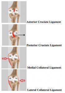
Four common ligaments of the knee are the Lateral Collateral Ligament (LCL), the Posterior Cruciate Ligament (PCL), and the two that are the most common to the public the Medial Collateral Ligament (MCL) and the Anterior Cruciate Ligament (ACL). The Patellar Ligament, referred to as the patellar tendon, is the ligament that connects the bottom of the knee cap (patella) and the top of the shin (tibia). Right above the patella is the quad tendon (quadriceps tendon) that connects the patella to the quads (muscles of the thigh). The meniscus is split into the medial and lateral aspects of the knee. These are cartilaginous “pillows” that rest on top of the tibia. The meniscus provides a cushion for the thigh bone (femur) to rest on the lower leg or shin.
What are the Collateral Ligaments of the Knee?
The LCL or Lateral Collateral Ligament and the MCL or Medial Collateral Ligament are on each side of the knee to help the side to side stability of the knee. The MCL attaches the femur to the tibia while the LCL attaches the femur to the fibula (the smaller, non-weight bearing bone on the outside of the lower leg).

What are the ACL and PCL Ligaments?
The quick answer is the ACL or Anterior Cruciate Ligament, and the PCL or Posterior Cruciate Ligament are fibrous structures that make an X in the center of the knee. Both are connected to the femur and the tibia. The PCL helps prevent the tibia from sliding backward when walking, running, etc. It goes from the back of the tibia to the front of the femur. The ACL helps prevent the tibia from sliding forward apart from the rest of the leg. It goes from the front of the tibia to the back of the femur.
The ACL is to be the most common injured ligament in the knee. The PCL has a better blood flow, and healing tends to be better.
If you want to learn more about the bones of the leg, this ARTICLE may help.
What are the Different Types of Knee Sprains?
- Grade I Sprain – This is where the ligament stretches but does not tear.
- II Sprain – When the ligament tears a little bit but does not completely tear.
- Grade III Sprain – This is a complete rupture of the ligament.

How are Ligament in Knee Injuries Diagnosed?
If you think you have a knee ligament injury, ask yourself these questions.
- Did you hear a pop when you got hurt?
- Is there swelling or maybe some discoloration of your knee?
- Does your knee feel unstable or loose?

Any or all of these are signs of knee ligament injuries. If you suspect that you have a knee ligament injury, then you should see an Orthopedic Surgeon. They will perform a physical exam and point you in the right direction if they think you need further testing. An X-ray will be the first test to rule out fractures (broken bones). The next test would be an MRI. This will look at soft tissue injuries such as ligament tears or meniscal injuries. After the medical professional has given you a diagnosis, they will set you up with a treatment plan. Some injuries are worse than others and require surgery. Watch this VIDEO on why Knee Pain Can’t Wait.
If you want to schedule an JOI Orthopaedic Knee Specialist appointment, please call 904-JOI-2000 or click below. JOI and JOI Rehab are Where The Pro’s Go!
Related Knee Articles:
- Finally, to schedule an appointment for physical or occupational therapy, or call 904-858-7045 or visit 12 area JOI Rehab Centers.