Watch This Video of Why Knee Pain Can’t Wait
How Do the Anatomy of Knee and Lower Leg affect Movement?
The knee is a hinge joint that sits between the thigh and the shin. It functions the same as a hinge on a door and sometimes gets a creaky as a hinge can. This joint allows the legs to bend and straighten, necessary for walking, going up and downstairs, going from sitting to standing, running, and jumping. The knee’s anatomy consists of many structures from the bones, tendons, and ligaments to the cartilage and muscles to help the knee function.
If you want to learn more about knee anatomy, please watch this knee anatomy video or this article Knee JOINT Anatomy.
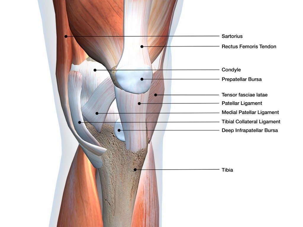 Anatomy of the Knee
Knee Anatomy: Bones and Joints
The anatomy of the knee consists of 3 main bones:
- The femur (thigh bone).
- The tibia (shin bone).
- The patella (knee cap).
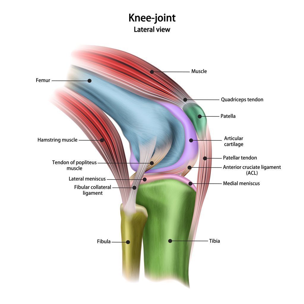 Lateral Knee Anatomy
The femur and the tibia are the main movers of the joint to allow for the hinge motion. This connection of the femur and tibia is a joint called the tibiofemoral joint. The patella sits on top of the tibiofemoral joint in a groove in the front of the femur. The patella is a floating bone that works as a fulcrum for the quadriceps muscle (you will read about this later) to function properly. This joint is called the patellofemoral joint and allows the patella to move up and down, and the knee bends and straightens.
Ligaments and Tendons of the Knee
The knee has 4 main ligaments:
- Medial collateral ligament (MCL): On the inside of the knee closer to the midline.
- Lateral collateral ligament (LCL): Is on the outside of the knee.
- An anterior cruciate ligament (ACL): Inside of the knee and crosses to the front.
- A posterior cruciate ligament (PCL): Inside of the knee and crosses to the back.
The MCL and the LCL sit on the sides of the knee, and they help give stability to the knee if your knee gets hit from the sides. 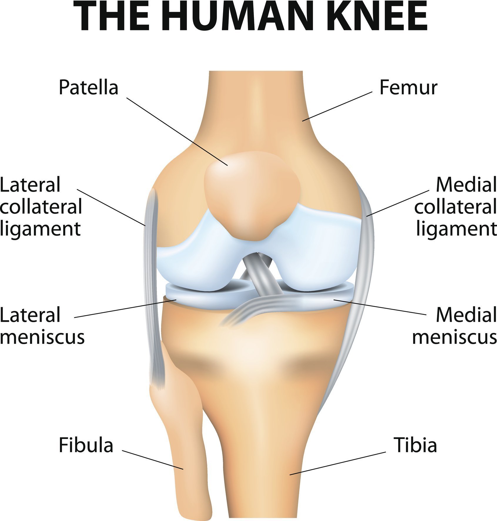 Knee bones, ligaments, and meniscus
The ACL and PCL are inside the knee and cross each other as they run front to back and vise versa. These 2 ligaments are responsible for giving the knee stability from front to back.
An ACL injury is probably one of the most recognized injuries in sports and, most of the time, requires a surgical repair that has a long recovery time. A full recovery after an ACL reconstruction is usually between 6 to 9 months depending on the patient and the other structures injured.
The unhappy triad is referred to when the ACL, MCL and Medial Meniscus are all injured at the same time. .
Tendons are where muscles attach to the bones of the knee. There are numerous tendons in the knee. The tendons which are prone to injuries of the knee are the Patellar Tendon and the Quadriceps Tendon. These patellar tendons can rupture or tear and they can also get tendonitis.
Cartilage of the Knee Joint
There are two main types of cartilage in knee anatomy: articular cartilage and the meniscus.
- Articular cartilage covers the bones’ ends and allows for the bones to slide and glide on each other without friction. This is the stuff you need to keep from getting the creaking and cracking of the joints. When this starts to wear down, arthritis will set in. Sometimes this cartilage is damaged with an ACL tear. The amount of trauma from the ACL injury can lesions to the cartilage of the joint or bones of the knee. This can be addressed during the surgical procedure.
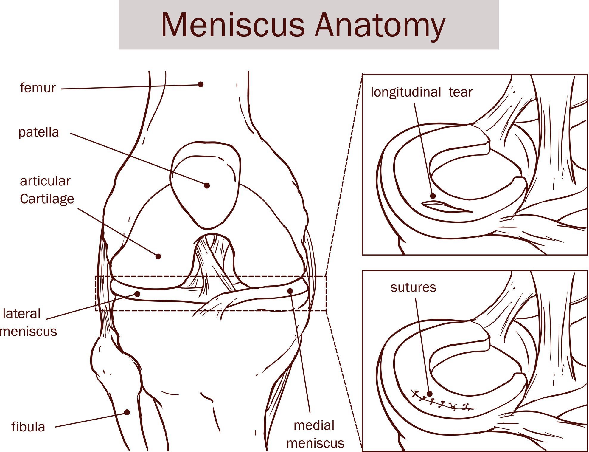 Image of articular cartilage and meniscus
- Meniscus: 2 thick pieces of cartilage that sit on the tibia between the femur and tibia. These are C-shaped that allow for improved congruence of the joint. Tears in these structures can cause pain, swelling, and sometimes catching and locking the knee joint. During surgery, the meniscus can be repaired or debrided. This is usually determined by the age of the patient, where the tear occurred and the amount of damage to the meniscus. To learn more, Read this Article about Meniscus Injuries.
Torn Ligament in the Knee
The quick answer is that a torn knee ligament can cause:
- Pain and the inability to walk
- Significant Swelling
- Feeling a “pop”
- Instability or loose feeling in the knee
- In sports, the athlete’s leg gives way during the sport.
To learn more, check out this article on Knee Ligaments.
Muscles and Tendons of the Knee
Many muscles affect the knee, but the main muscles that allow for the knee to perform its main functions are:
- Quadriceps: A group of 4 muscles that sits on the front of the thigh. These muscles are responsible for allowing the knee to straighten. This movement is necessary for standing from a seated position, bringing your leg forward when walking, and kicking a ball! The two patellar tendons attach the quad to the patella. These tendons can also rupture during sports.
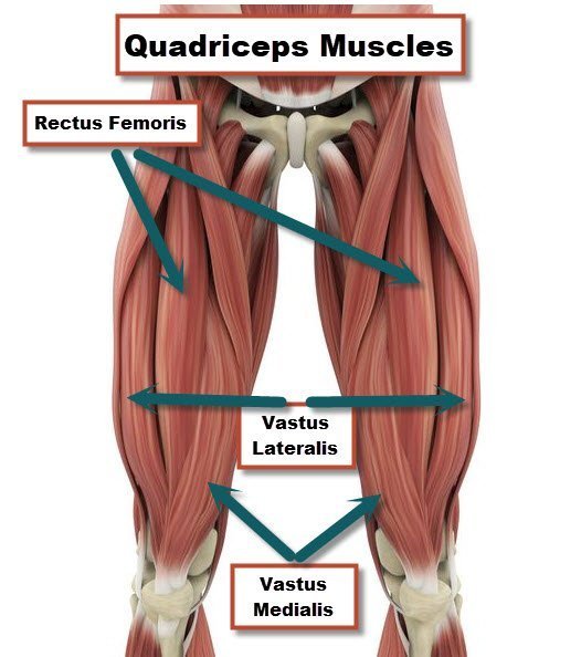 Quadriceps Muscle diagram
The Muscles in the Back of the Knee
- Hamstrings: A group of 3 muscles sits at the back of the thigh and allows for the knee to bend. These muscles are responsible for lifting your foot to walk. The hamstring muscles can be strained or torn during sport activities. The athlete is described by “pulling up” while running. This is a classic sign of a hamstring strain.
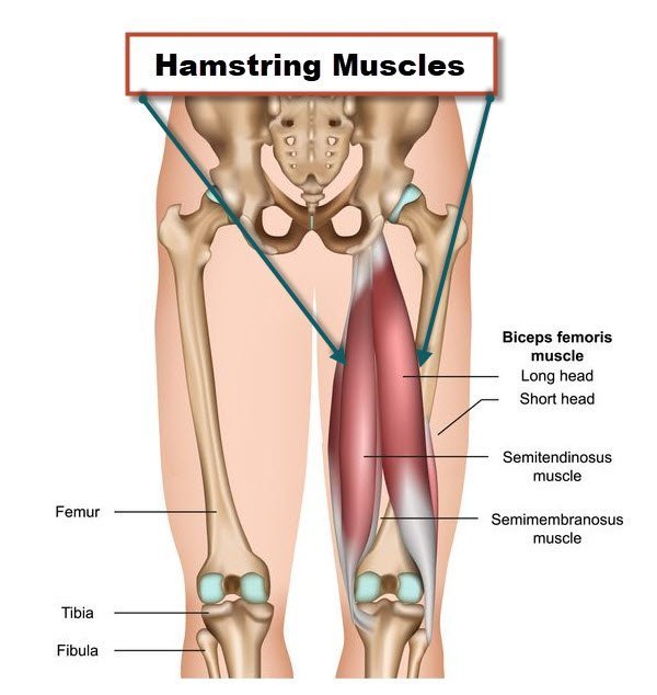 Hamstring Muscle diagram
- Gastrocs: A group of 2 muscles that sit in on the lower leg backside that works in tandem with the hamstrings to cause the knee to bend. The gastroc or calf muscle can be strained and torn during sports like tennis or basketball. The athlete will feel a “pop’ in the calf.
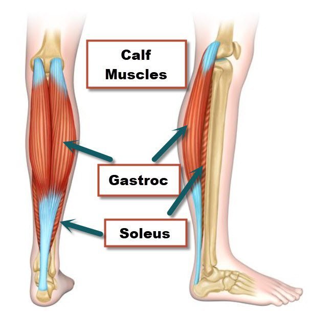 Calf Muscle Diagram
- Tendons attach the knee muscles to the bone. The two patellar tendons can also be prone to overuse and the development of patellar tendonitis. Jumper’s knee is common in the knee with athletics.
All of these muscles also have functions at different joints such as the hip and the ankle. Injuries to these structures, such as a pull or strain, will cause pain when activating the muscle and, if severe enough, will cause significant weakness.
Related Articles:
- Torn ACL
- Knee Ligaments
- What Does It Mean If My Knee Is Swollen?
- Best Low Impact Workouts for Knee Strength
Knee Doctors in Jacksonville
Many types of knee injuries can occur. Muscles, tendons, ligaments, and cartilage can be strained and sprained. It is really important to have your knee pain properly diagnosed by an orthopedic physician. JOI Rehab also has 12 Physical Therapy locations, which can certainly help you on the road to recovery. With over 90 Rehab Clinicians trained in providing you with the highest quality of orthopedic care. For an appointment, please call 904-858-7045.
To schedule an appointment for physical therapy at one of the 12 JOI Rehab Centers, please call 904-858-7045.
JOI MD’s now offer quick fracture care. Make an appointment by calling (904)JOI-2000, schedule online, or click the link below…