Tibia and Fibula Anatomy
The tibia and fibula are the two long bones of the lower leg. The tibia is the bone located on the inside of the lower leg, and the fibula is the bone located on the outside of the lower leg. Unlike the fibula, the tibia transmits most of the bodyweight during standing, walking, running, etc. These bones make up the bottom half of the knee joint and the top half of the ankle joint. To learn more about knee anatomy, please go to JOI Knee Anatomy.

How Serious is a Broken Tibia and Fibula?
The severity of a broken or fractured tibia is dependent upon what category of fracture that the injury belongs to. The two main categories of fractures are open and closed. Open fractures occur when the bone penetrates through the skin or if a wound is formed and the bone is visible. Closed fractures occur when the bone breaks, but the skin remains intact. All open fractures require surgery whereas casting may be recommended for a closed fracture. There are several types of fractures:
- Transverse: The fracture line is horizontal.
- Oblique: The fracture line is angled.
- Spiral: A fracture that resembles a spiral encircling the bone like stripes on a candy cane.
- Comminuted: Fracture breaking into 3 or more pieces.
Causes of Tibia and Fibula Fractures
Of the two bones, a fracture is more common in the tibia. Several causes can attribute to tibia and fibula fractures. Some include trauma from a high fall, contact sports, or vehicle accidents. Others are non-contact forces on the ankle, such as severe inversion (foot rolling in). Elderly individuals with osteoporosis can easily suffer fractures with falls or other mishaps.
Symptoms of Tibia and Fibular Fractures
Those with tibia and fibula fractures have severe pain at the location of the injury. Often there is a deformity present in the limb or a wound where the bone protrudes through the skin. If the fibula is only fractured, depending on severity, walking may be tolerable but likely very painful if it’s at the ankle level. With a tibial fracture, it’s highly unlikely the individual will be able to bear weight. Other symptoms include extreme tenderness, swelling, numbness due to artery or nerve damage, and bruising.
Diagnosis by X-ray
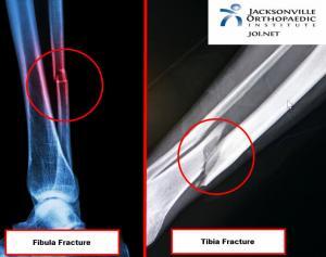
To confirm a fracture of the tibia and fibula, an X-ray is the first line of diagnostics and is usually sufficient to diagnose a fracture. Sometimes, stress fractures may not show up on an X-ray, and another form of imaging would be warranted. A CT scan will give a more in-depth image of the bone. An MRI will show the integrity of the ligaments, muscles, or any other soft tissue structures involved in the injury.
Treatment: Surgery vs. Non-Surgery
Can a Broken Tibia and Fibula Heal Without Surgery?
When a fracture is stable, a recommendation of casting without any surgical procedure is usually the best option. Usually, spiral and transverse fractures are stable enough for a cast. Oblique fractures are typically unstable and can shorten.
Surgery for a Tibia and Fibula Fracture
All open fractures will require surgery. Common surgeries consist of the following:
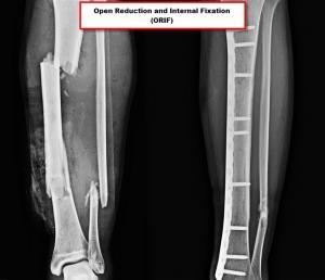
Open Reduction and Internal Fixation (ORIF): Consist of moving the broken bone fragments back into the original position, held in place by a metal plate held by screws.
Closed Reduction: The bone is realigned without making an incision at the site of injury.
Intramedullary Nailing: The placement of a rod into the medullary cavity of the bone with nails screwed to the 2 ends of the bone to stabilize the fracture.
External Fixation: Bone fragments are held in alignment and screws or pins support the leg from outside. For more severe and unstable fractures this procedure is the choice of the surgeon.
Potential Surgical Complications
- Blood clots.
- Fat embolism.
- Nerve damage.
- Infection.
- Malalignment of bones.
How Long Does It Take to Recover From a Fractured Tibia and Fibula?
Throughout the course of physical therapy, ankle mobility, strength, and balance improve. Swelling will decrease, and the patient is eventually able to weight bear and walk normally fully. The full recovery time of a tibia/fibula fracture takes between 3 and 6 months.
Physical Therapy after Tibia and Fibula Fractures
After surgery, physical therapy will begin as soon as the surgeon decides. As a result, the fracture or phase of the healing process will determine if a patient will either be non-weight bearing, partially weight-bearing/toe-touch weight-bearing, or full weight-bearing. It’s important to know that each surgeon’s protocol is individualized to their own restrictions based on the healing phase or surgery performed.
During the physical therapy evaluation, the physical therapist will likely find the following musculoskeletal impairments in the patient:
- Decreased ROM and strength in the ankle or foot.
- Decreased ability to put weight into the affected limb.
- Swelling.
- Atrophy.
- Decreased balance and proprioception.
Related Articles:
Ankle Arthroscopy: A Comprehensive Guide
Understanding turf toe injuries symptoms treatment
Preventing low ankle sprain tips and techniques
JOI and JOI Rehab
JOI Physicians continue to offer online new patient appointments. This is another option to make it more convenient to make new patient appointments with less phone hold times. Follow the link below to select your JOI MD and schedule online.
You can still call 904-JOI-2000 to make new patient JOI Physician Appointments if that is your preference.
To make appointments with JOI Rehab, please call 904-858-7045.