Category Archives: Uncategorized

JOI Uncategorized Archive

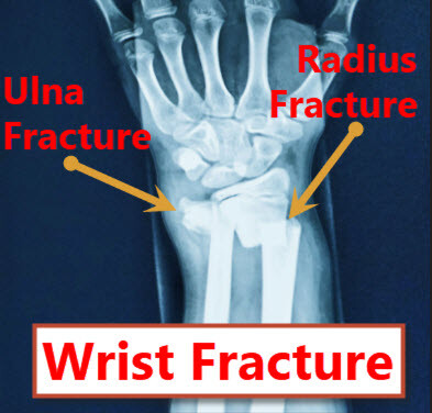
I Broke My Wrist

“I Broke My Wrist!” What Does This Mean?

Often in therapy we hear patients state, “I broke my wrist”. What exactly does that mean? In this article we will try to explain how to to diagnose and treat a broken wrist.

Overview of a Broken Wrist

The forearm is made up of two long bones: the radius and the ulna. The portion of the radius at the wrist level is known as the distal radius. A distal radius fracture (broken wrist) is the most commonly broken bone in the arm. The fracture usually occurs when a person falls on their outstretched hand. It can also happen in traumatic situations such a car accidents or during sports. Occasionally, the other bone in the forearm (the ulna) is also broken. When this happens, it is called a distal ulna fracture.

Wrist Hairline Fracture, I broke my wrist
Scaphoid bone fracture or broken wrist

Symptoms & Diagnosis

A distal radius fracture usually causes immediate pain, tenderness, bruising, and swelling. Occasionally, the wrist may look out of alignment if the fracture is severe. An X-ray of the wrist will be taken to visualize the extent of the injury. The fracture almost always occurs about 1 inch from the end of the radius bone (distal portion). A fracture that extends into the joint is called an intra-articular fracture. A fracture that does not extend into the joint is called an extra-articular fracture. The word “articular” means joint.

A fracture may also be displaced or nondisplaced. A displaced fracture occurs when the distal piece shifts out of alignment and results in deformity. A non-displaced fracture is when a fracture occurs with no change in alignment of the bone. A fracture that results in the bone breaking the skin is called an open fracture. A fracture in which the bone is broken into two or more pieces is called a comminuted fracture. Intra-articular fractures (fractures within the joints), open fractures (fractures that break through the skin), displaced fractures (distal portion shifts out of alignment), and comminuted fractures (fracture that shatter the bone into a lot of small pieces) are the more difficult cases to treat.

Non-surgical Treatment

If the broken bone is in a good position/alignment, a cast or splint may be applied until the bone heals. If the alignment of your bone is not good and likely to cause a functional deficit of the wrist and hand, it may be necessary to correct the deformity. In this case, the bone would be physically re-aligned (reduced) by the doctor. If the bone is straightened (reduced) without having to open the skin (incision), this is called a closed reduction. After the bone is properly aligned, a splint or cast may be placed on your arm. The cast may be changed as the swelling goes down so the cast does not get too loose.

Follow-up X-rays may be taken to determine that the bone is staying reduced and to ensure that proper healing is taking place. During this time, the patient may receive physical or occupational therapy to improve the motion in the uninvolved joints such as the fingers. When the bone is healed (usually about 6 weeks), additional therapy may be required to improve the motion and function of the injured wrist and forearm.

Distal radius fractures are the miost common wrist fractures to occur to the human wrist bone. JOI Rehab
Distal Radius Fracture

Surgical Treatment

If a distal radius has poor alignment and is not able to be properly reduced, a surgical treatment is usually warranted. If left untreated, long-term pain and deformity would prevent good functioning of the arm. There are many surgical options when dealing with a more complex distal radius fracture.

1. Percutaneous pinning (a pin inserted through the skin) may be used after the fracture is reduced to provide additional stability to the reduced fragments.

2. External fixation may be used to stabilize the unstable boney structure. In this kind of reduction, holes are drilled into uninjured areas of bones around the fracture and special bolts or wires are screwed into the holes. Outside the arm, a rod or a curved piece of metal with special ball-and-socket joints joins the bolts to make a rigid support. The fracture can be set in the proper alignment by adjusting the ball-and-socket joints. Since the bolts penetrate the skin, proper cleaning to prevent infection at the site of surgery must be performed.

3. Open reduction Internal Fixation (ORIF) may be used in which screws and/or plates are used to stabilize the fracture. Some surgeons use titanium bone plates, screws, and intramedullar nails to rigidly fix fracture.

After Surgery

In the early phases, ice, elevation (holding their arm up above their heart), compression garments to decrease swelling (ace wraps, isotoner gloves) and medications for pain relief are usually recommended. Due to the fracture being stabilized by the surgical hardware, therapy may be recommended days after surgery. Your physical or occupational therapist may work with you on pin care, custom splinting, swelling control, pain control, increasing mobility of the fingers, wrist, forearm, elbow, shoulder and neck and eventually strengthening when the fracture is healed enough.

Related Articles: Muscle Strain and Wide Awake Anesthesia.

JOI and JOI Rehab

JOI Physicians continue to offer online new patient appointments. This is another option to make it more convenient to make new patient appointments with less phone hold times. Follow the link below to select your JOI MD and schedule online.

You can still call 904-JOI-2000 to make new patient JOI Physician Appointments if that is your preference.

To make appointments with JOI Rehab, please call 904-858-7045.

Book An Appointment with a JOI Physician
Image of Book An Appointment with a JOI Physician

By: Tim Wall, MS, ATC

Groundbreaking Research from Bruce Steinberg, MD

Dr. Steinberg JOI REHAB
Dr. Bruce Steinberg

Jacksonville, FL, February 21, 2011 – Bruce Steinberg, MD, Jacksonville Orthopaedic Institute (JOI) surgeon, announces that his groundbreaking research paper, “Quantitative Muscle Hardness as a Non-invasive Means for Detecting Patients At Risk of Compartment Syndromes” is now in the publication by Physiological Measurement. Additionally, this is an international, peer-review publication. The paper co-authors were Ryan Riel, MD, Marshal Armitage, MD, and Hudson Berrey, MD, of the Department of Orthopaedic Surgery and Rehabilitation Health Science Center, Shands Jacksonville, University of Florida.

The publication is a journal for sensors, instrumentation and systems in physiology and medicine. It covers the quantitative assessment and visualization of physiological function in clinical research and practice. With an emphasis on the development of new methods of measurement and their validation.

Clinical Benchmark Study

Dr. Steinberg and his co-authors report on a benchmark clinical study validating quantitative muscle hardness and a device developed by Dr. Steinberg. In general, the device can be used to evaluate limb swelling, limb pain, muscle tone and muscle spasm. Also, the device can aid in the evaluation of medical conditions, such as limb edema, limb trauma, cellulitis, infections, hematoma, insect bites, snake bites, allergic reactions, contusions, stroke, cerebral palsy, paralysis, contractures, muscle strains and muscle ruptures.

There is demonstrated interest and excitement by the medical community for this device. It is non-invasive and shows great promise in providing additional diagnostic information for physicians thanks to Dr. Steinbergs groundbreaking research.

Make an Appointment Today!

Dr. Steinberg practices at the JOI San Marco physician office and Baptist Health Jacksonville.

Finally, to schedule with Dr. Steinberg, please call 904-564-2000 or schedule online or click below.

JOI & JOI Rehab Bold City Best 2022

Dr. Steinberg Press Release

Compartment syndrome can develop quickly in the limbs of patients with broken bones or crush-type injuries. The pressure of the swelling within the muscle compartment can increase enough to cut off the blood supply. Tissue death can occur within just six hours. The patient may permanently lose muscle function and even require amputation. Bruce Steinberg, MD, orthopaedic surgeon with Jacksonville Orthopaedic Institute, has spent the last 20 years developing an innovative tool to help physicians, nurses
and other medical personnel quickly screen whether a patient may have dangerous pressure elevation. The goal is to detect the rising pressure early enough to open the compartment and preserve the patient’s tissue. “In orthopaedics, compartment syndrome is a devastating problem if not detected and treated promptly,” Dr. Steinberg said.

Dr. Steinberg JOI REHAB
Dr. Bruce Steinberg

Physicians currently have two methods to make a diagnosis. They feel the area to determine its hardness (the area may feel swollen and tight, like it’s going to explode).The other method is a painful invasive test that involves placing a needle in the muscle compartment. Dr. Steinberg created and patented a noninvasive tool in which a spring-loaded plate is fitted around a 4-mm metal probe. The probe palpates against the muscle. The force of the pressure pushes the metal plate away, allowing a measurement of the muscle hardness. “It gives a quantitative measurement of palpation, from soft as a pillow to hard as a rock,” Dr. Steinberg said. The device is now in the testing stage. University of Florida (UF) residents used the device, along with other existing tools, to evaluate compartment syndrome in patients. Presentations on the data of its usefulness has been at three national meetings.

Scientific Paper Collaboration with UF

Most recently, Dr. Steinberg co-authored a paper about research on the device in collaboration with UF residents. This was under the direction of orthopaedic surgeon Hudson Berry, MD. The research was published by the peer reviewed international journal, Physiological Measurement, titled “Quantitative Muscle Hardness as a Noninvasive Means for Detecting Patients at Risk for Compartment Syndromes.” (April 2011, Vol. 32, No. 4, pp 433-434). A large clinical trial is now underway testing multi-trauma patients at Orlando Regional Medical Center.

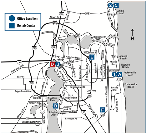
New Year’s Insurance Changes

Update Your Health Insurance Information!

It’s that time again… time to let JOI and JOI Rehab know of any New Year’s Insurance Changes. If you are a patient with JOI or JOI Rehabilitation, here’s what you need to do before you are seen in 2023:

  1. Bring us any New Health Insurance Cards
  2. Inform our Front Desk Staff of any Changes in your health insurance coverage for January 2023
  3. Bring any new ID cards with updated address or contact information.

Other Insurance Changes

Be aware that many health insurance plans have Deductibles and out of Pocket Maximum Expense limits that will start over on January 1. Copayment amounts may also change depending on your insurance plan. We recommend that you check with your insurance carrier about any potential changes. If you have any questions about the new year’s insurance changes, give us call and we can help!

COVID-19 in 2023

JOI and JOI Rehabilitation will continue to follow CDC guidelines for protecting you against COVID-19 in 2023. Temperature screenings and Universal Mask Mandates will continue at all JOI and JOI Rehabilitation facilities.

We look forward to meeting the challenges of 2023 with you! Let’s get you on the Road to Recovery!

To schedule an appointment, call 904-JOI-2000 or schedule online.

JOI and Jeff Lageman

Jeff Lageman

Jeff Lageman is the featured celebrity for the Jacksonville Orthopaedic Institute Super Bowl Commercial. Lageman is a former first round draft pick in the National Football League. He played football in high school for the Park View High Patriots. He was both linebacker and defensive end before playing at the collegiate level for the University of Virginia Cavaliers. Lageman was taken 14th overall by the New York Jets in 1989. He played in New York until 1995 when he was signed by the Jacksonville Jaguars during their inaugural year. Lageman played a total of 10 years in the NFL. He finished his career with a total of 47 sacks. He then moved on to the next phase of his professional career in sports by becoming a broadcaster.

Jeff Lageman Radio Broadcaster

Due to his love for hunting, fishing, and the outdoors Lageman began a radio show in Jacksonville called “The Outdoors Show”. In 2014 Lageman became a team member of the official radio broadcast of the Jacksonville Jaguars and is still active in that role today. Lageman also appears on several other radio shows on the network 1010XL.

JOI Super Bowl Commercial

This is the 5th straight year JOI has aired a Super Bowl commercial and couldn’t be more proud of its work in the local athletic community. JOI is the Official Sports Medicine Provider for the Jacksonville Jaguars as well as other professional teams. With 26 ATC’s, JOI is in professional sports, but in the local high school and college community as well. JOI provides sports medicine services to Florida State College of Jacksonville and Jacksonville University as well as 7 high schools: Atlantic Coast, Bishop John J. Snyder, Bishop Kenny, The Bolles School, The Episcopal School, Paxon, and Fernandina Beach High School. Jacksonville Orthopedic Institute has designed a vertically integrated network for athletes in all aspects of the recovery process. JOI has Orthopedic Sports Medicine Physicians, Physical Therapists, Physiatrists, 12 Physical Therapy Centers and a dedicated Sports Center, among several other services all under one roof. This unique network of professionals provides unmatched care that is individually tailored to get you back on the road to recovery.

Why JOI?

Jacksonville Orthopaedic Institute is committed to providing the best care for its patients, and in the words of Jeff Lageman “I want to go where the best is, and me, I’m going to JOI”. JOI, Where the Pro’s Go!

Finally for more information visit JOIonline.net or call 904-JOI-2000.

Skip to content