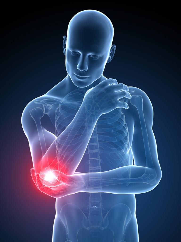
Although no surgeon or patient ever wants a patient to need a joint replacement revision, especially with most joint replacements having a 90% success rate, they are sometimes necessary. Failed joint replacements can occur for a variety of reasons, including wear and tear or complications that develop over time.
If you’ve had a joint replaced, it’s important to be aware of potential joint failure symptoms. Recognizing a failed joint replacement early could mean an easier revision surgery and fewer post-op healing complications. When it’s time for a joint replacement revision, being prepared will give you both the knowledge of what to expect and peace of mind, so you don’t fear the unknown.
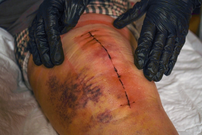
A revision surgery differs from the primary surgery because the conditions are different — the surgeon isn’t dealing with an untouched problem that needs to be fixed. In addition to the primary issue, there may be other complicating factors that must be addressed. For example, scar tissue may have formed around the site, making the surgery more complex.
Unfortunately, revision joint surgeries are often more complicated because there can be bone loss, scar tissue, and carry higher risks for infection and instability. Recovery can take longer. While joint replacements have come a long way, revision joint surgery may be required for those who have had knee, shoulder, or hip replacements, among others.
Joint replacement revision is different than a primary joint replacement surgery because everything is just a bit more complicated. The original implant can present unique challenges, as well as additional changes to the joint and anatomy post-surgery that the surgeon must account for when revising.
Common Reasons Joint Replacements Fail
If a joint replacement revision is needed, it’s often a result of one of the following reasons:
Implant Loosening
Over time, your joint implant may shift or become misaligned, causing pain, discomfort, or instability. If it impacts your mobility or causes you pain, it may be time for a revision, but your provider will determine the best course of action for your specific case.
Infection
Although infection can be treated with medication, it can require a revision surgery. Infections can create a biofilm that attaches to bone and the implant, impacting functionality, and a complication that antibiotics alone cannot remove. Signs of an infected joint replacement can include redness, warmth to the touch, and a fever. Signs of an infection require prompt treatment to prevent it from advancing.
Implant Wear and Component Failure
Especially if a joint surgery was completed many years ago with outdated prosthetic materials and technology, the prosthetic material can break down over time. When the component wears out and fails, it is time for a revision joint surgery. Those who are more active may be at risk for the joints wearing out sooner.
How Doctors Diagnose Failed Joint Replacements
If you’re concerned that your joint replacement has failed, a licensed orthopedic specialist will conduct a thorough review to determine whether a revision is necessary. The steps typically include a physical examination and labs.
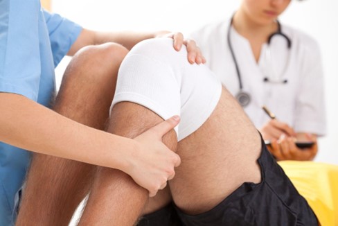
The physical examination will include a detailed account of your symptoms, as well as a physical examination in which your doctor will manipulate the joint to assess functionality, including instability, reduced mobility, and more. Your doctor will carefully examine your joint for signs of infection, such as redness, warmth, and swelling.
Additionally, you will likely be sent for additional testing beyond the physical examination, which will help support your physician’s diagnosis. Your doctor will likely order imaging tests, which could include X-rays, CT scans, and MRIs. Lab testing will likely also take place to analyze joint fluid, as well as blood tests to look for infection.
Treatment Options Before Revision Surgery
If you’re concerned you might need a joint replacement revision, it is important to explore treatment options before jumping into another surgery. Surgery is hard on the body, and recovery after a revision will likely take longer than the initial surgery. Non-surgical treatment options are often used in combination to achieve the best possible outcome.
Some options to explore before deeming your joint a failed joint replacement include physical therapy to improve strength and stability. Working with your provider on pain management options, including medications or injections, that can help you feel a little better while working on physical therapy, for example. Additionally, you may need temporary activity modifications to reduce stress on your joint.
Tips for Protecting Your Joint Replacement
If you’ve had joint replacement surgery and are concerned you may need a revision in the future, there are some things you can do to be proactive about maintaining your primary replacement.
Patients must attend follow-up appointments and follow the post-op instructions, including required and recommended physical therapy. This can reduce the risk of abnormal healing or scar tissue formation.
Additionally, patients should do their best to maintain a healthy weight. Being overweight puts additional wear and tear on joints, including pressure that can ultimately cause an injury over time. Staying physically active while being cognizant of the replacement (doing your best to prevent injuries and falls) will go a long way in protecting your joint replacement.
Contact an Orthopedic Specialist
If you’re experiencing symptoms that might indicate your joint replacement isn’t working as it should, it’s best to get it evaluated, especially if you’ve been doing well but are now experiencing problems. Symptoms that shouldn’t be ignored include instability or a feeling that your joint is “giving out,” swelling and pain, noises or other strange sensations, stiffness, and decreased mobility. It’s important to have your joint evaluated by a qualified professional, as early intervention can lessen the chances of scar tissue setting in or of other complications from popping up. Contact a qualified orthopedic professional like the physicians at the Jacksonville Orthopaedic Institute today to get an appointment to have your joint evaluated!
If you’re like 25% of adults who suffer from chronic knee pain, you’re likely looking for relief. This common, sometimes debilitating condition often comes as a result of injuries, overuse, and simply aging. While surgery can be a recommended treatment option for some patients, other non-surgical options for chronic knee pain treatment can lead to better outcomes or the same outcome as surgery.
Exploring non-surgical options before seeking a surgeon may give you many years of pain-free health and movement without having to go under the knife. Chronic knee pain treatment can come in many forms, from knee injections to other options for orthopedic care.
Understanding Chronic Knee Pain
Knee pain is very common, but it doesn’t always affect people the same way. Knee pain can be a result of a variety of different conditions, including knee arthritis and injuries, among others. Understanding chronic knee pain, what causes it, and options for chronic knee pain treatment will best prepare those with the pain to make decisions.
What Is Chronic Knee Pain?
Chronic knee pain is defined as knee pain that persists for three months or longer, with the most common cause being osteoarthritis. Chronic knee pain is pain that does not subside. It may get worse or aggravated from time to time, or it may stay at the same level of pain or discomfort, but to be considered chronic, it must continue for months.
Common Causes
There are a number of causes for chronic knee pain. Some may result from an injury, overuse, or even just a progression of aging. Some common causes include:
Osteoarthritis
Tendonitis
Meniscus injuries
Ligament strain
Obesity and joint stress
Benefits of Non-Surgical Treatment Options
When considering your next steps for chronic knee pain treatment, it is wise to consider the benefits of non-surgical treatment options. While you may end up having surgery at some point, knowing that you exhausted non-surgical options is worth peace of mind and financial savings.
Non-surgical treatment options often carry much lower risk than surgery, even if that turns out to be warranted. Recovery isn’t linear, and it isn’t the same for every patient. Surgery requires significant recovery time, time off work, and may involve potential complications. The patient will also need to strictly adhere to rehab schedules to realize the benefit of the surgery, and not doing so could jeopardize the surgery’s success. There is an inherent risk that not everything will go to plan.
Additionally, surgery is expensive, and if there are complications that require a hospital stay or multiple return visits, the cost only goes up. The financial burden can be immense if a patient has a high deductible or insurance that doesn’t completely cover the treatment.
The overall benefits of at least trying non-surgical chronic knee pain treatment are significant, as it could mean complete healing without surgery. Personalized treatment plans can help improve mobility and manage or eliminate pain.
Non-Surgical Chronic Knee Pain Treatment Options
With the guidance of a physician specializing in orthopedics, along with physical therapists, there are a variety of options to treat knee pain, even knee arthritis, without surgery.
Physical Therapy and Exercise
Many instances of chronic knee pain can benefit from muscle development to help support the knee. A physical therapist can help prescribe a plan to increase strength in the muscles that support the knee, improve mobility and flexibility, and increase the range of motion.
Strengthening Exercises
Strengthening exercises and weight training may be indicated if an individual has insufficient support for their joint. By improving the support muscles surrounding the knee, normal knee function may be possible, which relieves pain and improves mobility.
Stretching and Flexibility
Many people are often quite stiff, especially if they are sedentary. Improving range of motion through stretching exercises and increasing flexibility may be options that can help relieve knee pain.
Weight Management and Lifestyle Changes
Unfortunately, many individuals experience knee pain because they are overweight. Obesity and even simply carrying a few extra pounds can result in reduced pressure on the knee joints. This can allow them to move freely without impeding the range of motion.
Weight management and lifestyle changes, including adding low-impact exercises, can help the patient lose weight and reduce the pressure on those joints. Consulting with a nutritionist can help patients make healthier choices, and in some cases, an anti-inflammatory diet may be recommended.
Low-Impact Activities
Staying active is another component of weight management and overall health, in addition to helping with chronic knee pain treatment. An orthopedic physician and/or physical therapist may recommend that a patient engage in the following low-impact activities that can help get patients moving while not putting extra pressure on their joints.
Swimming
Cycling
Walking
Yoga
Medications and Pain Relief Options
In some cases, a multi-faceted approach that includes adding certain medications and pain relief options is necessary. Your doctor will know which medications can help you without causing further problems down the road.
Over-the-Counter Medications
Some pain relief medications can be purchased over the counter, but those are indicated for acute situations, not chronic problems. Those with chronic knee pain should seek guidance from licensed physicians specializing in orthopedic care.
Topical Treatments
The market for topical treatments, including creams and gels, has expanded, and there are plenty of options that can provide some immediate, localized relief. These will have analgesic properties but won’t do anything to address the source of the problem.
Prescription Medications
Prescription medications are those medications that cannot be purchased at the local pharmacy without a prescription. For example, some prescription NSAIDs are better for long-term use because they don’t cause as much GI upset as those available without a prescription. Of course, there are also stronger pain medications that must be used with caution and under the direction of a physician.
Injection Therapies
Injection therapies are also a great option for chronic knee pain treatment. Knee injections can provide short-term benefits, including anti-inflammatory relief and increased lubrication, and there is some evidence that platelet-rich plasma therapy (PRP) can help regenerate tissue.
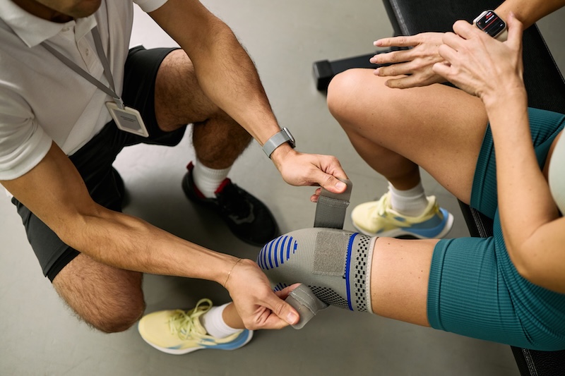
Bracing and Supportive Devices
In some cases, bracing and supportive devices may be all a patient needs to get relief, or a combination of medications, knee injections, and bracing. These mobility aids could include knee braces and sleeves, orthotics and shoe inserts, and other mobility aids.
Find Out Your Options for Non-Surgical Chronic Knee Pain Treatment
Every case is different and should be evaluated by a medical professional, especially if you’re experiencing worsening symptoms, instability, or persistent swelling. There are many options to treat chronic knee pain, and it’s important to determine the best one for your specific condition. That’s where the Jacksonville Orthopaedic Institute comes into play. Our team of doctors and physical therapists can provide you with a professional evaluation that can maximize long-term joint health and promote overall well-being without being rushed into surgery. Contact Jacksonville Orthopaedic Institute to schedule your orthopedic care evaluation today.
If you’ve been told you might need joint replacement surgery, you may be wondering how long do joint replacements last. Similarly, if you’ve already had a joint replaced in the past and are potentially experiencing old symptoms coming back, you may want to know what the joint implant lifespan is or if you’ll ever need to have a surgical revision.
There is a lot to consider and review when it comes to joint replacement surgery, whether it’s your first or you’ve already had one. Joint replacement surgery involves a lot, though in some cases, recovery may not be as bad as the individual expects. However, you will need time to recover, physical therapy to get moving again after surgery, and it is important to factor in, if applicable, how you may heal from a second surgery in the same location.
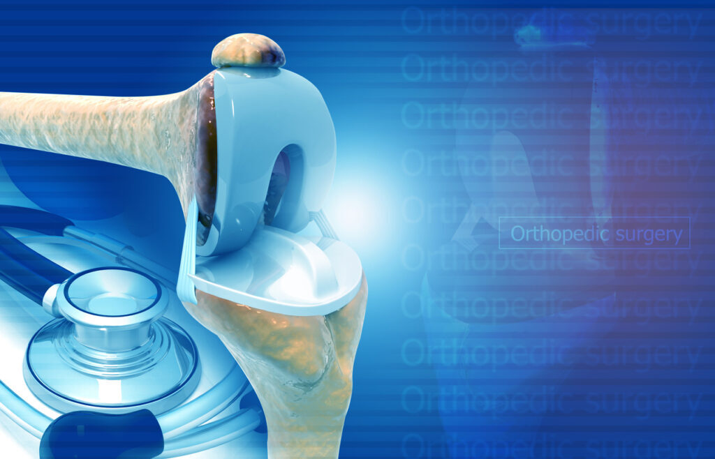
Joint replacement surgery, also known as arthroplasty, is a procedure that removes damaged, diseased, or otherwise incompatible joints and replaces them with artificial joints. The artificial joints, called prostheses, are often made of metal (such as titanium), ceramic, or even plastic. Commonly replaced joints include hips, knees, and even shoulders.
Average Lifespan of Joint Replacements
Joint replacements are very common surgeries, despite being major undertakings that require extensive recovery and care. For the most part, joint replacements are expected to last a decade or more, depending on the type of joint replaced. The American College of Rheumatology estimates that joint replacement for hips and knees lasts 25 years or longer, with most being a lifelong solution. Rheumatology is the study of rheumatic diseases, like arthritis, that affect the joints and surrounding tissues.
Harvard Medical School reports that there are 600,000 knee replacements and about 330,000 hip replacements each year, with both lasting 10 to 15 years. With that many surgeries, there are bound to be anomalies, meaning that although there may be an average joint implant lifespan, it is critical to acknowledge and understand that every case is unique.
Just because there is an average, it does not mean that it is guaranteed to be the case for you. You may have better-than-average results, or you may end up being the individual whose surgery isn’t as successful as others. Your doctor will help guide you and help you have a realistic expectation of how your surgery will go and whether you may need further surgeries down the road.
Factors That Affect Longevity
There are many factors that affect how long joint replacements last. Ultimately, success depends on the individual person. For each individual, their age at the time of surgery, their health, their weight, and even their activity level before and after surgery can have a major impact on how well the surgery goes, how they recover, and, of course, how long the implant lasts.
In addition to the patient, the surgeon also affects the lifespan of the joint implant. The initial placement and success of the surgery will, in part, dictate how everything else goes. If there are problems immediately, it may negatively affect the outcome, and vice versa. Additionally, the type of implant material used and even the surgeon’s techniques can dictate success. Needless to say, the conditions that can affect the surgical outcome are vast and vary widely among patients.
Other factors that affect how long do joint implants really last include access to healthcare and rehabilitation, as well as compliance with rehabilitation requirements. Access and compliance with rehabilitation have a major impact. A person may have access to the best rehab facilities with the most advanced rehabilitation plan, but if they aren’t compliant, it will negatively affect their outcome. Conversely, if they don’t have access but are willing, they may not be able to achieve the same results.
Signs a Joint Replacement May Be Wearing Out
If you’ve had a joint replacement surgery and are concerned your joint implant’s lifespan may be nearing the end, you will want to contact a qualified orthopedic specialist. Symptoms of joint issues returning, such as increased pain and stiffness, reduced mobility or stability, and swelling or redness, mean you need to get in to see a medical professional.
Unfortunately, many of the old symptoms returning could mean you may be experiencing a complication with your joint. While this might mean you need a revision surgery to change the implant or adjust its placement, outcomes can still be positive. Revision timelines vary depending on what needs to be done. Either way, if you’re having issues, you should see your surgeon or consult another expert in the field of orthopedics to figure out what’s going on.
Tips to Help Your Joint Implant Last Longer
If you’ve recently had a joint replacement and are trying to figure out how long do joint replacements really last, the good news is that there are things you can do to be proactive about increasing the longevity of your joint implant lifespan.
Your joints carry your weight, so it is imperative that you maintain a healthy weight. This does not mean you should go to extreme measures or adopt a diet to be as small as possible, but it does mean you are at an appropriate weight for your height and muscle mass. Being purposeful about fitting fitness into your life after recovery is important to living a long, healthy life in general, and it also benefits your joints.
Always follow your surgeon’s post-surgical advice, prescribed physical therapy, and recovery guidelines as they are intended to help you heal in the best way possible. Maintain regular checkups with your doctor and do your best to stay in good health because it helps all of you, including your joint implant!
Get Expert Advice From Jacksonville Orthopaedic Institute
Before you go under the knife, it’s important to be proactive and get a qualified opinion from a licensed healthcare provider who can personalize your health improvement plan, whether it’s with a joint revision surgery, initial joint replacement surgery, or even a non-surgical option for pain relief. The team of highly trained and specialized orthopedic physicians, surgeons, and physical therapists at Jacksonville Orthopaedic Institute is here to help you. Contact us today for a consultation.
If you’re like many people who have never endured a surgery and are experiencing chronic knee pain, you’re likely stressed, worried, and trying to figure out a knee pain treatment without surgery. Although there are several common causes for chronic knee pain, some of which may require surgical treatment, it is important to realize that there are effective knee pain treatments without surgery.
Knee pain treatment without surgery can include a variety of modalities, from injections to physical therapy for your knee and other non-surgical options. By finding a provider who will advocate for non-surgical treatments and exhaust all options before turning to surgery, you may be able to find relief from your chronic knee pain.
Understanding Common Causes of Chronic Knee Pain
It is important to understand the source of your knee pain before you and a trusted healthcare practitioner can determine if there is a knee pain treatment without surgery you can try. Chronic knee pain, meaning it persists and doesn’t go away, and isn’t the result of a traumatic injury, can be caused by changes to your bones, tendons, ligaments, or even a result of simple overuse over time. No one wants to go through surgery unless it’s necessary, and some conditions can be healed without it. With that said, it’s always best to be evaluated by a qualified orthopedic doctor to know for sure.
1. Osteoarthritis
Osteoarthritis is a common cause of chronic knee pain that stems from degenerative changes to the joint tissue. The joint tissue helps keep your joints, including your knees, operating smoothly without noise (like popping, squeaking, or creaking), pain, or strange sensations, like stiffness. Because this condition is degenerative, it unfortunately means it will get worse over time — and it can negatively affect the other components of a joint, such as your tendons, ligaments, and meniscus. While surgery may end up being necessary at some point, it is not the first line of defense against chronic knee pain.
2. Tendinitis, Overuse, and Strains
Additionally, tendinitis, overuse, or repetitive strain can cause knee pain that persists. Tendinitis occurs when there is swelling in your tendons, which can be caused by an injury, like a strain, or from overuse, such as the case with jumper’s knee, a sports-related injury. Often, knee pain treatment without surgery is the preferred course of action for these chronic pain issues.
3. Ligament Injuries and Meniscus Tears
Ligament injuries and minor meniscus tears may heal over time without surgical intervention. Ligaments are the connective tissue that connect the two points of your joints together, providing stability while they move. Your meniscus is a type of cartilage that acts almost like a shock absorber in a vehicle, preventing pain, abnormal movement, and stopping bone-on-bone interaction, which is painful.
For those with minor ligament injuries and meniscus tears, there is the option for knee pain treatment without surgery, though a doctor may recommend surgery if the tear or injury is severe or traumatic. These are common causes of knee pain that can improve without surgery.
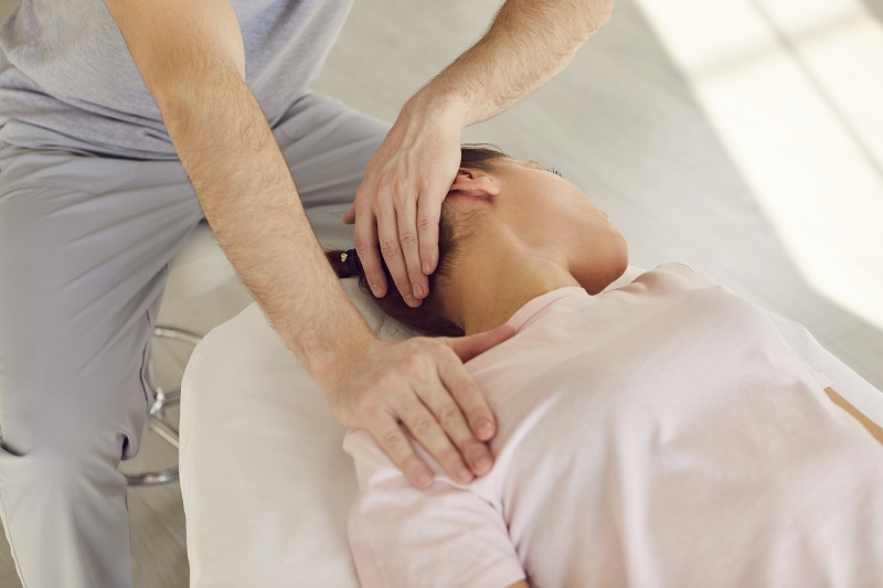
Physical Therapy And Exercise
You may be wondering how you can heal the various causes of chronic knee pain, and what exactly you can do to get knee pain treatment without surgery. Physical therapy for knees is often a great solution for healing that pain, providing both improved stability, reduced or eliminated pain, and tools for success moving forward. A physical therapist is a licensed medical professional trained specifically to diagnose and treat movement injuries, diseases, and other conditions, including chronic pain.
Physical therapy for knees can provide the patient with knee pain treatment without surgery, depending on the case, of course, and frequently it is the first line of treatment for injuries. Physical therapy can provide the patient with much-needed pain relief through guided stretching and low-impact strengthening exercises that won’t harm the joint, and it can also provide the knowledge and tools to prevent future injury.
The physical therapist may also recommend supportive tools or assistive devices, such as braces, canes, walkers, and orthotics, to help manage pain and provide much-needed joint stability and support.
Medications For Pain Relief
Pain management is a major component of knee pain treatment without surgery. If an individual can effectively manage their pain and continue their normal activities of daily living without hindrance, they may not need surgery!
Pain relief is often used in conjunction with physical therapy, lifestyle changes, and additional support. Pain-relief medications can come in different forms, including over-the-counter oral medications, prescription medications, topical creams and gels, and injections. Supplements, like glucosamine, chondroitin, and MSM, may also help.
Over-the-counter options for pain relief include anti-inflammatory medications (NSAIDs like ibuprofen) and other analgesic-type medications like acetaminophen. It is very important to know that long-term consumption of over-the-counter pain medications can cause damage to your internal organs. Safe pain management for knee pain treatment without surgery should still be overseen by a medical professional.
Prescription oral pain medications may also be used to treat both chronic pain and acute symptoms. However, keep in mind that these must be prescribed by a doctor and carefully monitored to ensure there are no adverse effects or resulting addictions.
Additionally, injections, such as a corticosteroid shot, must be administered by a licensed healthcare professional, often only in a healthcare facility. Topical pain relief gels and creams could include pain relievers, which would be prescribed. However, some over-the-counter options offer cooling and analgesic effects.
Lifestyle Changes That Make a Difference
Knee pain treatment without surgery often includes elements of physical therapy, pain management, and lifestyle changes. Lifestyle changes can have a major positive impact on an individual’s life, the pain they experience, and their mobility.
Ensuring an individual has proper footwear and support, and proper bracing if needed during activity, can make a difference in how much a person can move without pain. Additionally, activity modification may mean cycling instead of playing basketball or walking instead of running.
Rest and recovery are important components of non-surgical treatments for chronic knee pain that many overlook. An individual’s weight is also a common contributing factor to joint pain that must be addressed. Ultimately, joint stress reduction is a must.
Alternative And Complementary Therapies
Anyone who is carrying extra pounds likely already knows that their weight can affect how their joints feel, but they may not know how to lose weight to feel a difference. Having a multifaceted approach to knee pain treatment without surgery means pulling together different resources, like physical therapy, a nutritionist or registered dietician, and proper mental support, like with a counselor or therapist, that can all work together to solve the problem. Lifestyle changes are often critical in successfully treating knee pain without surgery.
Additional alternative and complementary therapies that could be introduced into an individual’s lifestyle and have a positive effect include acupuncture, massage therapy, and chiropractic care. Some holistic medicine practices can offer benefits, though there is often insufficient published research to support them either way. It is important to consult with medical professionals who offer a range of orthopedic care options to find the best fit for your condition and preference.
Contact Jacksonville Orthopaedic Institute
If you’re experiencing chronic knee pain and want help, contact the Jacksonville Orthopaedic Institute. Our team of caring, multifaceted professionals offers a wide range of personalized treatment options designed to get you living without pain —including knee pain treatment without surgery. Many people are able to find relief without having to go under the knife. Schedule a consultation today to see how we can help address your knee pain.
At some point or another, a person is likely to feel hip pain. Whether a person over-exercised or is just getting older, it is a fairly common ailment. However, in cases where the pain persists or other symptoms accompany the pain, it’s important not to ignore it. Understanding why it’s bothering you and knowing what caused the pain will go a long way in determining the appropriate treatment protocol.
Often, hip pain is due to one of two main conditions: osteoarthritis or sciatica. Both conditions cause pain, may lead to diminished mobility, and can have long-term problems if left untreated. Osteoarthritis and sciatica can even occur simultaneously. Having a qualified physician examine you and determine the cause of your discomfort and pain is essential for creating a treatment plan that can relieve your hip pain.
Understanding Osteoarthritis of the Hip
Osteoarthritis is a degenerative joint disease that happens when cartilage inside the joint begins to break down. This can happen as we age or as a result of untreated trauma to the area that affects the cartilage. Those with osteoarthritis may also experience pain in other joints, in addition to hip pain, as it is a joint disease.
When osteoarthritis affects the hip, the cartilage, which is the smooth tissue that covers the hip bone (medically known as the femoral head), begins to degrade. That cartilage helps the hip bone glide inside the pelvis, specifically in the acetabulum or the socket. When it can’t move smoothly, you feel pain.
Who is at Risk for Osteoarthritis?
People most at risk for osteoarthritis are older adults, more commonly women over 50. Those who are overweight put more stress on their joints, which can lead to both osteoarthritis and sciatica. Additionally, individuals who have suffered a past injury are involved in sports that can aggravate the hip joints or that simply require a lot of use. Some individuals may have a genetic predisposition to osteoarthritis or have joints that don’t line up properly, which can cause the condition to flourish.
Common Symptoms of Hip Osteoarthritis
If you are concerned you might have osteoarthritis or sciatica, you may be experiencing pain that simply isn’t going away. Both conditions cause persistent hip pain and discomfort. However, some unique features point to osteoarthritis over sciatica.
With hip osteoarthritis, you may experience pain that gets worse when moving, stiffness that is often worse in the morning or after sitting for long periods, and reduced range of motion. You could have hip osteoarthritis. Additionally, you may be experiencing a clicking noise or grinding sensation coming from that joint. Your pain will likely get worse with weight-bearing activity.
Osteoarthritis and sciatica have a lot of overlapping symptoms, but osteoarthritis has a gradual onset that leads to more pain, limited mobility, and further issues over time. Sciatica is a nerve pain, so the pain associated with sciatica doesn’t necessarily grow or change.
Understanding Sciatica
While hip pain is often related to the joint, as in osteoarthritis, sciatica is pain that stems from the sciatic nerve. The sciatic nerve runs from the base of the spine down the leg. It is often related to back conditions, such as disc herniation or spinal stenosis, or even an injury like a pelvic fracture. Individuals feel pain when the nerve is impacted, whether by a disc pressing on it or the bones in the spine narrowing and pressing on the nerve. Either way, the nerve becomes irritated and inflamed.
Common Symptoms of Sciatica
Sciatica has unique symptoms that are unlike osteoarthritis, though you may feel a general sense of pain or discomfort in the hip area, which can lead to confusion about the source of the pain. Sciatica is a nerve pain that is the result of an irritated or compressed sciatic nerve. It typically does not feel like the dull ache or stiffness that individuals experience with osteoarthritis. Sciatica instead often feels like a sharp, burning, or searing pain that often travels through the buttocks and down the leg. You may experience tingling or numbness in addition to the pain, as well as weakness, and prolonged pain with sitting or standing.
Key Differences Between Osteoarthritis and Sciatica
While osteoarthritis and sciatica have unique pain indicators, some overlapping pain patterns can prove to be confusing for laypeople. By considering where exactly you’re experiencing pain, the kind of pain it is, what makes it worse, and how it came about, you may be able to have a better idea as to whether you’re dealing with osteoarthritis or sciatica.
Location of Pain
With osteoarthritis, pain is deep within the groin or the front of the hip; it is pain that is emanating from the hip socket as the cartilage that allows for smooth movement has become damaged. Sciatica pain often starts in the back, or at the very least, the back of the hip, and radiates through the buttock and down the leg.
Type of Pain
While sciatica pain is often described as sharp, burning, and shooting, osteoarthritis pain is not described in that manner. Unlike sciatica, osteoarthritis pain is stiff, mechanical, and can be described as a dull ache.
What Makes It Worse
Considering what exacerbates pain is a big indicator in determining whether you are suffering from osteoarthritis and sciatica or one or the other. Osteoarthritis is aggravated by walking and bearing weight on the joint, whereas sciatica is aggravated by sitting or standing for prolonged periods.
How did it Start
Additionally, how the hip pain started is a key difference between osteoarthritis and sciatica. Hip pain that begins gradually and gets worse over time is more often linked to osteoarthritis. Conversely, sciatica is a nerve pain that starts suddenly and stays at the same level. It can come on suddenly or after an injury.
Get Your Hips Checked For Osteoarthritis and Sciatica
If your pain is not going away and you’re concerned that you may be suffering from osteoarthritis or sciatica, it’s time to get checked out. Add in increasing limitations, progressive stiffness, numbness, or new symptoms like weakness appearing, and it’s likely there might be a problem. Of course, if there are any emergent symptoms, like loss of bowel or bladder, you need to head to the emergency room. There are some key differences between the two conditions, but you can leave the diagnosis to the experts at the Jacksonville Orthopaedic Institute. While both conditions are treatable, early intervention and evaluation always lead to a better prognosis. Contact us at JOI today — we’re here to help you and get pain relief from hip pain!
Everyone has joints, and as we age, we often notice that we don’t bounce back as quickly as we used to, or that round of golf starts to produce some aches and pains that weren’t there last year. If your joints have been causing pain and you’ve noticed it happening more frequently, persistently, or with increased severity, you could be suffering from joint damage.
Joint damage symptoms start subtly, but as the condition deteriorates, everything gets worse, which is why early intervention is the best course of action. Early detection can prevent joint damage symptoms from going off the rails, impacting your mobility and quality of life. With early detection, your doctor can create a treatment plan that can help with pain prevention and management, as well as slowing the progression of certain joint disease conditions. Joint damage symptoms can arise from a variety of reasons, including autoimmune diseases, injury, and osteoarthritis, among other painful conditions.
What Is Joint Damage?
Joint damage occurs when the cartilage, which is the smooth tissue that surrounds the end of the bone in a joint, begins to break down. That breakdown can cause inflammation and irritation, impacting how your joint normally functions. Joint damage symptoms can result from structural changes in the joint, causing joint noises, increased pain, stiffness, and other problems, ultimately affecting how your joints function.
When you’re active, you may occasionally experience soreness in your joints, especially from overuse. This can commonly happen in the elbows of people who play a lot of tennis or golf. However, temporary soreness gets better with rest, while progressive damage continues to cause issues, stiffness, pain, clicking sounds, and more, indicating a larger problem with the cartilage, not just a one-time irritation.
Early Warning Signs You Shouldn’t Ignore
While feeling a twinge of pain that goes away with an over-the-counter anti-inflammatory, ice, and rest isn’t a problem, some early warning signs might not seem so bad at first, but they are symptoms of a larger problem. The adage “an ounce of prevention is worth a pound of cure” is accurate in many scenarios. By paying attention to early warning signs, you can help prevent further damage. Pay attention to your body and the signals it is sending you, as they may lead to serious injury if ignored. Here are some early warning joint damage symptoms you should heed:
1. Persistent Joint Stiffness
If you’re experiencing joint stiffness, especially in the morning or after sitting, it might be a sign of joint damage. If the stiffness persists after you get moving and warm up, it may also point to a bigger problem than a one-off, minor issue.
2. Swelling Around the Joint
If you notice your joint is swollen, that’s a warning sign that something is wrong. Inflammation is visible and can be felt as puffiness, swelling, and warmth at the joint. This level of inflammation is a red flag that your joint damage symptoms warrant evaluation by a healthcare professional.
3. Mild but Recurring Pain
If you’re experiencing constant pain, pain that lasts longer and more frequently, or that just keeps coming back despite periods of rest and home treatment, it’s a sign that you need to pay attention to your body. Aching is another sign that something is going on with your joint.
4. Reduced Range of Motion, Weakness, or Instability
Other red flag joint damage symptoms include difficulty bending, straightening, or moving your joint — or more plainly put, your joints might not feel like they’re working normally or the way they have in the past. If you find your mobility is limited or affected in any way, you need to see a doctor. Your health and quality of life depend on it!
5. Clicking, Popping, or Grinding
Sometimes it’s normal for our joints to make occasional cracking sounds. However, regular or persistent clicking, popping, or grinding may indicate deterioration of joint cartilage. Joint damage symptoms that signal cartilage wear need to be evaluated.
Who Is at Higher Risk for Experiencing Joint Damage Symptoms?
Certain populations will experience joint damage symptoms more than others. Those at higher risk for these types of ailments include older adults over the age of 50, those with a history of joint injuries, and those with a family history of arthritis. Additionally, those who are overweight put more stress on their joints, which means they are at a higher risk of developing joint problems. Athletes and those who repeatedly bend and use their joints at work may also be at a higher risk of developing joint damage symptoms.
Why Early Treatment Matters
You might be thinking, if my joints are shot, there’s nothing they can do! While this may be true in a worst-case scenario, it is often not the case for most patients when the disease is treated early. Early treatment can improve the prognosis and slow the progression of joint damage, making your joint damage symptoms more manageable for longer.
Additionally, by seeing a doctor at the first sign of concern rather than ignoring problems, you can potentially prevent a permanent joint deformity and avoid surgical intervention or joint replacement. Overall, having your joint damage symptoms evaluated early can improve the long-term quality of your life and help you manage discomfort more easily, maintaining your mobility for longer.
Get Your Joint Damage Symptoms Evaluated
If you’re concerned about your joints or experiencing early warning joint damage symptoms, it is important to be proactive about your health. Even if it turns out to be a minor irritation and not a symptom of long-lasting joint damage, being proactive about your health means you are positioning yourself to protect your joints and preserve joint health now and in the future.
Schedule an appointment with one of the esteemed physicians at Jacksonville Orthopaedic Institute today — you won’t regret taking care of your health!
Joint replacement surgeries are often described as a life-changing procedure. Whether it’s the knee, hip, or shoulder, most people go into surgery expecting pain relief, improved mobility, and a return to their normal activities. When pain lingers months after your surgery, it may make you feel exhausted from options and have questions.
Is pain after joint replacement normal? Or is ongoing discomfort a sign that something is wrong?
The answer to these questions depends on several factors, such as how long the pain lasts, how intense it is, and the type of pain.
Understanding the Healing Timeline After Joint Replacement
Before determining whether the pain is normal or a concern, it is important to understand the timeline of your joint replacement surgery. These procedures can involve cutting through bone, muscle, and connective tissue. Even though damaged joint surfaces are replaced, your body still needs to heal after undergoing this procedure.
Early Recovery: 1-6 Weeks
During the first couple of weeks, pain is common and should be expected. This pain can come from:
Surgical trauma to tissues.
Swelling or inflammation.
Muscle weakness and stiffness.
Increase in activity due to physical therapy.
Pain during this period typically improves, although there may be a few flare-ups after rehabilitation sessions.
Intermediate Recovery: 6 Weeks-3 Months
By this time period, you may notice significant improvement. Pain levels are discrete, swelling subsides, and your mobility and range of motion increase. Mild soreness or stiffness is normal, especially after activity, but it should be manageable.
Long-Term Recovery: 3 Months- 1 year
Some discomfort may persist for months after the procedure, as the muscle strengthens and the joint adapts to the implant. However, pain should be minimal and not interfere with your daily life. If your pain is worsening, there may be an underlying issue.
What is Considered Normal Pain After Joint Replacement?
Normal pain after joint replacement may exhibit specific characteristics. Understanding these can help determine whether the pain is due to healing or to potential issues.
Normal Pain:
Improves gradually over time.
Occurs after activity and improves with rest.
Feels like soreness, stiffness, or muscle ache.
Responds to medication, ice, or physical therapy.
It is also normal to experience these symptoms while healing:
Morning stiffness.
Temporary pain flare-ups.
Mild swelling or warmth around the joint.
As you heal, these symptoms should gradually decrease, not increase.
When Ongoing Pain is Not Normal
While discomfort is expected, ongoing or worsening symptoms should not be ignored, as this can indicate an underlying issue.
Red Flags
Pain that worsens.
Sharp, stabbing, or burning sensations.
Pain that continues during rest or sleep.
Significant swelling, redness, or warmth.
Limited movement that doesn’t improve despite physical therapy.
Pain with chills or fever.
Common Cases of Joint Pain After Surgery
There are many reasons you may experience pain after joint replacement surgery.
Inflammation and Soft Tissue Irritation
Muscles, tendons, and ligaments surrounding the joint may be irritated for a few months following surgery. Scar tissue can also contribute to stiffness and discomfort.
Nerve-Related Pain
Nerves can be stretched or irritated during surgery, leading to burning or tingling sensations, numbness, or radiating pain down the limb. Nerve pain typically feels different from joint pain and can take longer to resolve.
Muscle Weakness
Muscles around the joint can weaken quickly after surgery. If these muscles don’t regain their strength, it can lead to abnormal movements and ongoing pain.
Overuse
Depending on the patient, postoperative activity levels may vary. Your physical therapist will recommend the amount of movement you should perform to reduce pain. Overdoing it too soon can cause pain and delay healing, making it essential to follow the recovery plan.
Implant Problems: Pain Signaling a Bigger Issue
While some may experience pain due to minor issues like inflammation or irritation, others may be caused by a more serious problem. Persistent pain may indicate an implant problem:
Common Implant Issues
Implant Loosening: Over time, your implant may loosen from the bone, causing deep, persistent pain, a feeling of instability, and pain during weight-bearing activities.
Implant Misalignment: If the implant is positioned incorrectly, it can cause stress and lead to chronic discomfort or abnormal movement.
Wear and Tear: Implants can experience wear and tear, especially in younger, active patients. This can irritate the surrounding bone and soft tissue.
Allergic Reactions: Some patients may react to metals used in the implant, resulting in inflammation and pain without infection.
Infection Around the Implant
Although less common, infection is a serious complication. Here are some symptoms to look out for:
Persistent pain.
Swelling and Redness.
Drainage near the surgical site.
Fever or fatigue.
Infections can occur after surgery or years later.
When Should You Seek Medical Attention?
You should contact your healthcare team if:
Pain continues after three months.
Pain interferes with sleep or daily activities.
Your symptoms are new or worsening.
You suspect an infection or implant instability.
Treatment Options for Persistent Implant Pain
Treatment options will depend on what is the underlying cause of the pain.
Conservative Treatments:
Physical Therapy adjustments.
Anti-inflammatory medications.
Pain management.
Activity modifications.
Interventional Options:
Corticosteroid injections.
Nerve-targeted treatments.
Treatment for underlying spine or muscle issues.
Surgical Intervention:
If there is a problem with the implant, like instability or infection, surgery may be necessary.
How Doctors Evaluate Ongoing Pain after Joint Replacement
Your healthcare team may use several tools, such as tests, studies, and examinations, to determine the root cause of pain. They may perform:
A physical examination to look at movement, stability, and tenderness.
Imaging tests like X-rays, CT scans, or MRIs.
Blood tests to check for infection.
Joint aspiration to analyze fluid near the implant.
Frequently Asked Questions
How long should pain last after joint replacement? Most patients experience gradual improvement over 3–6 months, although mild soreness may persist for up to 1 year.
Is severe pain months after surgery normal? Severe or worsening pain months later is not typical and may indicate implant problems or infection.
Can implant loosening cause pain years later? Yes. Implant loosening can develop over time and cause deep joint pain and instability.
When should I worry about joint pain after surgery? If pain interferes with sleep, worsens over time, or is accompanied by fever or swelling, contact your surgeon.
Jacksonville Orthopaedic Institute
If you are experiencing pain after your joint replacement surgery, contact the Jacksonville Orthopaedic Institute today. JOI offers physicians who specialize in joint replacement surgery. JOI continues to offer online new-patient appointments as an additional option to enhance convenience, with shorter phone hold times. Follow the link to schedule online with a JOI physician.
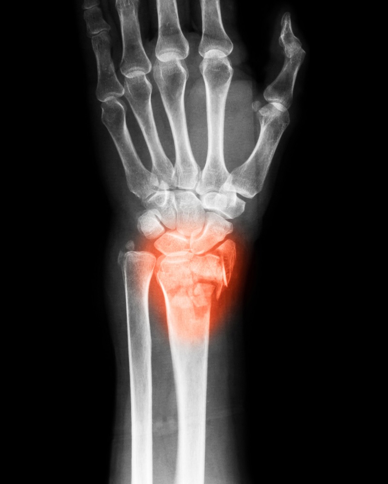
If you have been experiencing discomfort or pain in your wrist or hands, you are not alone. Many people experience wrist pain, but understanding its causes and distinguishing between carpal tunnel and arthritis is key to finding the right treatment plan for your condition. Though both can lead to similar symptoms, carpal tunnel and arthritis have two distinct causes and treatments.
What is Carpal Tunnel Syndrome?
Carpal Tunnel Syndrome (CTS) is a condition that affects the median nerve that runs down the forearm, all the way to the palm of your hand. The nerve passes through the carpal tunnel, a narrow passageway of ligaments and bone located in each wrist. When this tunnel is compressed, the median nerve can be pinched, causing discomfort, pain, tingling, numbness, and weakness in the hand and wrist. This is often caused by representative hand movements or swelling.
Common Symptoms of Carpal Tunnel Syndrome
Here are a few common symptoms that can help differentiate carpal tunnel vs. arthritis:
Tingling or numbness in the thumbs, index, middle, and part of the ring finger.
Pain that worsens at night, especially after prolonged wrist use.
Weakness in the hand that makes gripping difficult.
Swelling and pain that improve with massaging or shaking the hand.
Nerve compression symptoms that worsen with repetitive activities like typing or using a mouse
Risk Factors for Carpal Tunnel Syndrome
Several factors can lead to an increase in the likelihood of developing CTS:
Repetitive wrist movements typically result from daily activities such as typing.
Pregnancy can lead to fluid retention, putting more pressure on the median nerve.
Obesity can contribute to inflammation and compression in the carpal tunnel.
Underlying health conditions, such as diabetes, can increase the risk of nerve damage.
How is Carpal Tunnel Diagnosed?
Your healthcare provider may begin by performing a physical examination, assessing your symptoms, and performing tests (such as the Phalen’s test or Tinel’s sign) to check for nerve compression. In some cases, other studies, such as electromyography (EMG) or nerve conduction studies, may be ordered to confirm the diagnosis or assess severity.
What is Arthritis?
Arthritis is a broad spectrum of conditions that cause joint inflammation and pain in or near the joints. There are more than 100 types of arthritis, but the most common are rheumatoid arthritis (RA) and osteoarthritis (OA).
Both types of arthritis can affect the wrist joints, leading to joint inflammation, discomfort, and pain. Symptoms are often confused with CTS.
Types of Arthritis That Affect The Wrist
Rheumatoid Arthritis (RA): An autoimmune condition in which the immune system attacks the joints, causing joint inflammation, pain, and, in severe cases, deformity.
Osteoarthritis (OA): Unlike RA, osteoarthritis is a degenerative joint disease characterized by the breakdown of joint cartilage over time. OA can affect the wrist, leading to stiffness, pain, and reduced range of motion.
Post-Traumatic Arthritis: This condition develops after a wrist injury, resulting in chronic joint pain and stiffness.
Common Symptoms of Arthritis in the Wrist
Arthritis in the wrist is primarily characterized by joint inflammation, unlike CTS. Here are a few key symptoms:
Pain in the wrist that worsens after prolonged activity.
Swelling or visible deformities near the joint.
Stiffness and limited wrist mobility make it difficult to bend or twist.
Warmth around the joint, typically seen in RA cases.
Morning stiffness that lasts for more than 30 minutes.
Risk Factors for Arthritis in the Wrist
Arthritis may be more likely depending on:
Age: As you get older, osteoarthritis becomes more common.
Genetics, especially with rheumatoid arthritis.
Joint injuries or a previous trauma can lead to arthritis later down the line.
Gender, as women are more likely to develop arthritis.
Obesity can put strain on the joints.
How is Arthritis Diagnosed?
Arthritis can be diagnosed through a physical exam and imaging studies like X-rays or MRI scans. Blood tests can help confirm the type of arthritis by detecting antibodies.
Carpal Tunnel VS. Arthritis: Key Differences
While both conditions can cause wrist pain, their symptoms, causes, and treatments differ. Here’s how to tell the two apart:
Location of Pain
Carpal Tunnel: The pain, tingling, and numbness associated with CTS typically affects the palms, thumb, index finger, and middle finger.
Arthritis: On the other hand, arthritis affects the joints themselves. In the wrist, this means the pain is typically over the joint and may radiate up the forearm or down to the hand.
Symptom Timing
Carpal Tunnel: The pain in CTS often worsens at night and can wake you up from sleep. It also tends to flare up after repetitive wrist activities.
Arthritis: Arthritis pain is usually more constant, especially if joint inflammation is present. Morning stiffness is also a common symptom in those with this condition. Pain may increase after overuse or periods of inactivity.
Type of Pain
Carpal Tunnel: CTS pain is typically described as sharp or burning and may also cause tingling or numbness due to nerve compression.
Arthritis: Pain from arthritis is typically more achy or dull and is often accompanied by joint stiffness.
Joint Changes
Carpal Tunnel: In general, there are no visible changes to the joint when CTS is present. CTS is more nerve compression rather than inflammation or damage to the joint itself.
Arthritis: It can cause visible joint changes, such as swelling, deformities, or reduced range of motion. In severe, long-term cases, joint damage or bony growths may be seen on imaging studies.
Carpal Tunnel vs Arthritis: Quick Comparison
Feature
Carpal Tunnel Syndrome
Arthritis
Cause
Median nerve compression
Joint inflammation or cartilage breakdown
Pain Type
Burning, tingling, numbness
Achy, stiff, swollen
Location
Thumb, index, middle fingers
Wrist joint itself
Worse At
Night, repetitive activity
Morning, after inactivity
Visible Swelling
Rare
Common
Diagnostic Tests
EMG, nerve conduction study
X-ray, MRI, blood tests
Jacksonville Orthopaedic Institute
If you have symptoms similar to carpal tunnel or arthritis, contact Jacksonville Orthopaedic Institute today. JOI offers physicians who specialize in CTS and arthritis conditions. JOI continues to offer online new-patient appointments as an additional convenience, with shorter phone hold times. Follow the link to schedule online with a JOI physician.
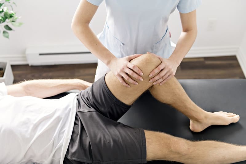
An Anterior Cruciate Ligament (ACL) injury is often a major setback for athletes and active individuals. Once the pain subsides or surgery is complete, ACL injury prevention becomes a key focus of the recovery process. Preventing re-injury is one of the most critical and challenging goals, and physical therapy plays a vital role. Whether the injury occurred through sports, exercise, or an unexpected incident, recovery doesn’t end once pain has subsided or surgery is complete.
This is why physical therapy plays a major role in ACL injury prevention. Physical therapy will help you restore strength, movement control, and confidence while reducing the risk of a second injury.
Understanding ACL Injuries and Re-Injury Risk
The ACL is one of the main ligaments that stabilize the knee joint. It helps maintain forward movement and stability, especially during cutting, pivoting, and jumping, which are common in most sports and exercises.
After an ACL injury, the knee is left vulnerable. Research has shown that individuals who return to sports and exercise too soon or without proper rehabilitation face a significantly higher risk of re-injury.
A second injury often occurs because:
Strength has not been restored fully.
Movement patterns are incorrect.
Compromised knee stability.
Loss of neuromuscular control.
Physical therapy addresses all of these factors, tailored to your case, to ensure knee stability before returning to normal activity.
Why Physical Therapy is Essential for ACL Injury Prevention
Physical therapy focuses on both healing and prevention. A rehab program will focus on rebuilding the knee’s ability to withstand stress safely.
Key goals of physical therapy for ACL injury prevention include:
Restoring strength and muscle.
Improving knee stability.
Correcting form and movement mechanics.
Enhancing balance with coordination.
Without physical therapy, these elements may not be restored to their full potential, increasing the risk of re-injury, especially for athletes.
Restoring Strength Through ACL Rehab Exercises
One of the most common causes of ACL reinjury is muscle weakness. After an ACL tear, the quadriceps, hamstrings, and hip muscles weaken due to inactivity, swelling, and altered movement patterns.
Targeted Muscle Strengthening
Physical therapists design ACL rehabilitation exercises to restore lower-body strength. Here are a few examples of exercises you may do at physical therapy:
Quadriceps strengthening for knee control.
Hamstring exercises to protect the ACL.
Glute and hip strengthening to reduce knee strain.
Working different muscle groups will help stabilize the knee during dynamic movements and reduce stress on the ligament.
A key advantage of physical therapy is its ability to progress. At the beginning, you may be heavily limited, but as you progress through the program, exercises gradually increase in difficulty to ensure knee stability and muscle strengthening, helping you return fully to activities.
Improving Knee Stability and Joint Control
Knee stability is more than just ligament strength; it also includes the joint, muscles, and nervous system.
Proprioception and Balance Training
Proprioception is the body’s ability to sense joint position, and after an ACL injury, it is often impaired. Physical therapy will focus on balance and coordination to restore this system.
Common techniques:
Single-leg balance drills.
Stability training (on uneven surfaces).
Controlled landing and deceleration exercises.
As proprioception improves, knee stability will increase, reducing movements that could lead to ACL reinjury.
Neuromuscular Training
Neuromuscular training teaches the body to move more efficiently and safely. This is essential for preventing ACL injuries, especially among athletes.
By retraining movement patterns, physical therapy reduces mechanics that commonly lead to ACL tears.
Correcting Faulty Movement Patterns
Many ACL injuries and re-injuries are from faulty movement patterns.
High-Risk Movements
Physical therapists closely analyze how you:
Jump and land.
Pivot and cut.
Accelerate and decelerate.
Improper knee alignment, excessive knee collapse, or poor control can place stress on the ACL. Physical therapists aim to help patients develop safer movement patterns to prevent re-injury.
Retraining and Athletic Movements
Corrections are applied not only to sports movements but also to everyday activities such as walking, stair climbing, and lifting.
The Role of Sports Therapy
Sports therapy is a specialized version of physical therapy that focuses on athletic performance and injury prevention. Sports therapy is often the final, if not the most crucial, phase of rehabilitation after an ACL injury.
Sports-Specific Conditioning
Sports therapy programs create the physical demands of the patient’s specific sport. This can include:
Agility drills.
Plyometric training.
Sprinting and cutting exercises.
Reaction-based movement drills.
These activities will prepare the knee for real sport and in-game scenarios once you return to full play.
Gradual Return-to-Play Progression
Structured progression is one of the most beneficial aspects of sports therapy. Athletes are guided step by step, reducing the likelihood of injury from returning to sports activities too soon.
Mental Readiness and Confidence Building
Just like with any recovery from an injury, many patients experience fear of re-injury after an ACL tear. Even if the knee is physically strong and ready to return to activity, hesitation or lack of confidence can still affect movement.
Physical therapy helps with mental readiness by:
Rebuilding trust in the knee.
Gradually exposing patients to challenging movements.
Reinforcing correct mechanics through repetition.
Confidence building and mental readiness are just as important as the physical side of therapy.
Long-Term Benefits of Physical Therapy
After rehab ends, the benefits of physical therapy will continue.
Individuals who complete a full ACL rehabilitation program experience:
Improved knee stability.
Improved athletic performance.
Reduced chances of re-injury.
Increased confidence in movement patterns.
Many therapists provide a maintenance program for patients who have completed the ACL rehab exercises, which they can complete independently.
Because patients with ACL injuries are at higher risk of injuring the opposite knee, physical therapists also work to prevent ACL injury in the opposite leg by addressing whole-body mechanics, correcting asymmetries, and training both legs symmetrically.
Jacksonville Orthopaedic Institute
JOI Physicians continues to offer online new-patient appointments. Our team of experienced orthopedic knee specialists will work with you to develop a customized treatment plan tailored to your specific needs and goals. Follow the link to select your JOI MD and schedule online.
Rotator cuff symptoms can start subtly, but they’re one of the most common causes of persistent shoulder pain. Shoulder discomfort and pain can be hard to ignore, especially since we use our shoulders in everyday activities. Whether you are having trouble lifting your arm, shoulder instability, or weakness during movement, or persistent pain even while relaxing, there may be a deeper problem. One of the most common causes is a rotator cuff injury. Recognizing rotator cuff tear symptoms early can significantly impact treatment plans, recovery, and long-term shoulder health.
Understanding The Rotator Cuff
The rotator cuff consists of four muscles and tendons surrounding the shoulder joint. Together, these muscles and tendons stabilize the shoulder, enabling smooth movement when lifting, rotating, or reaching. Because of the shoulder’s wide range of motion, the joint relies heavily on the rotator cuff for both strength and control.
Why Early Detection Matters
Since symptoms can start subtly, most people delay treatment. Untreated rotator cuff injuries can worsen, leading to chronic pain, reduced mobility, muscle weakness, and possibly loss of function. Identifying rotator cuff tear symptoms early will give you the best chance at successful treatment and recovery. The earlier rotator cuff tears are detected, the greater the likelihood of less invasive treatment with better long-term outcomes.
What Are The Early Signs of a Rotator Cuff Tear?
Early symptoms of rotator cuff tears are often mistaken for general shoulder strain. Here are a few common symptoms of rotator cuff injury:
Persistent Shoulder Pain
One of the most common rotator cuff tear symptoms is continuous shoulder pain that doesn’t improve with rest. Many describe this pain as:
Dull or aching
Worsens at night, especially if lying on the injured side.
Increases with lifting.
This type of shoulder pain tends to persist and gradually worsen over time.
Weakness in the Shoulder or Arm
Difficulty while lifting or rotating the arm is another warning sign for injury. Many notice weakness when:
Reaching overhead
Lifting objects away from the body.
Performing everyday tasks that require lifting, like combing hair or reaching a shelf.
This weakness typically develops slowly, making it easy for people to adapt to it rather than look for the underlying cause.
Limited Range of Motion
Stiffness or reduced mobility may indicate a tendon injury. With a limited range of motion, you might struggle to:
Raise your arm.
Reach behind your back.
Perform circular motions.
Losing your range of motion while experiencing pain is often a key indicator of a potential rotator cuff tear.
Clicking or Popping
Some people experience grinding, clicking, or popping near the joint when moving the shoulder. While it may not be painful, it can be an indication of irritation to the tendons or damage within the joint.
Increasing Pain
Early rotator cuff tears typically begin with mild discomfort that gradually worsens and becomes painful. Pain may start off only during activities, but over time can happen even while resting.
How Do You Know If a Rotator Cuff Tear Has Worsened?
If left untreated, symptoms may worsen or become more disruptive to your daily life.
Severe and Constant Pain
If the pain has become sharper, more intense, and constant, affecting not only when moving, but during sleep as well, you may have an advanced rotator cuff tear. Shoulder pain at night and while sleeping is a common sign of more progressed tears.
Significant Decrease in Strength
Shoulder strength may decline significantly as the tear enlarges. Simple activities can become increasingly more difficult.
Inability to Lift Arm
In severe cases, some people experience “pseudoparalysis” that makes lifting their arm without assistance impossible. If you experience this symptom, schedule an appointment for an orthopedic exam as soon as possible.
What Causes a Rotator Cuff Tear?
Age-Related Degeneration
As we age, the rotator cuff tendons weaken and lose elasticity. Degenerative tears are most common in adults over 40 and can develop without injury.
Repetitive Activity
Jobs or sports that involve repetitive motions, such as painting, swimming, and baseball, can increase the risk of tendon wear and tear.
Acute Injury
An acute injury can occur from something as simple as lifting a heavy object or having a fall. A sudden trauma can lead to an immediate tear that is often accompanied by pain and weakness.
Poor Posture
Poor posture and weak shoulder stabilizers can place excessive stress on shoulder muscles and tendons, increasing the risk of injury.
When Should You See A Doctor for Rotator Cuff Pain?
Certain rotator cuff tear symptoms should prompt an orthopedic exam by your physician.
You should seek medical attention if you experience:
Persistent shoulder pain
Pain that worsens even while at rest.
Weakness or loss of function.
Difficulty sleeping due to shoulder pain.
Seeking treatment at the first sign increases the likelihood of successful treatment and positive long-term outcomes.
What to Expect During an Orthopedic Exam
An orthopedic exam is necessary for diagnosing a rotator cuff injury. During the exam, your physician will:
Review your medical history and create a timeline of your symptoms.
Assess shoulder strength, function, and range of motion.
Perform physical tests to evaluate tendon function.
Imaging studies like X-rays, ultrasounds, or MRIs may be ordered to confirm the diagnosis and help determine treatment options.
How Are Rotator Cuff Tears Treated?
Non-Surgical
Early or partial tears can respond well to conservative options like:
Activity changes
Anti-inflammatory medication
Physical Therapy
Corticosteroid injections
This approach aims to reduce pain, improve function, and prevent further injury.
Surgical
Surgery may be recommended for those who have:
Full and advanced tears
Severe weakness and loss of function
Symptoms do not improve despite conservative treatment.
Jacksonville Orthopaedic Institute
If you experience any of the early rotator cuff tear symptoms, do not ignore them; early medical intervention will increase your chances of a positive, successful outcome.
JOI Physicians continue to offer online new patient appointments. Our team of experienced orthopedic shoulder specialists will work with you to develop a customized treatment plan tailored to your specific needs and goals. Follow the link to select your JOI MD and schedule online.